Samsung
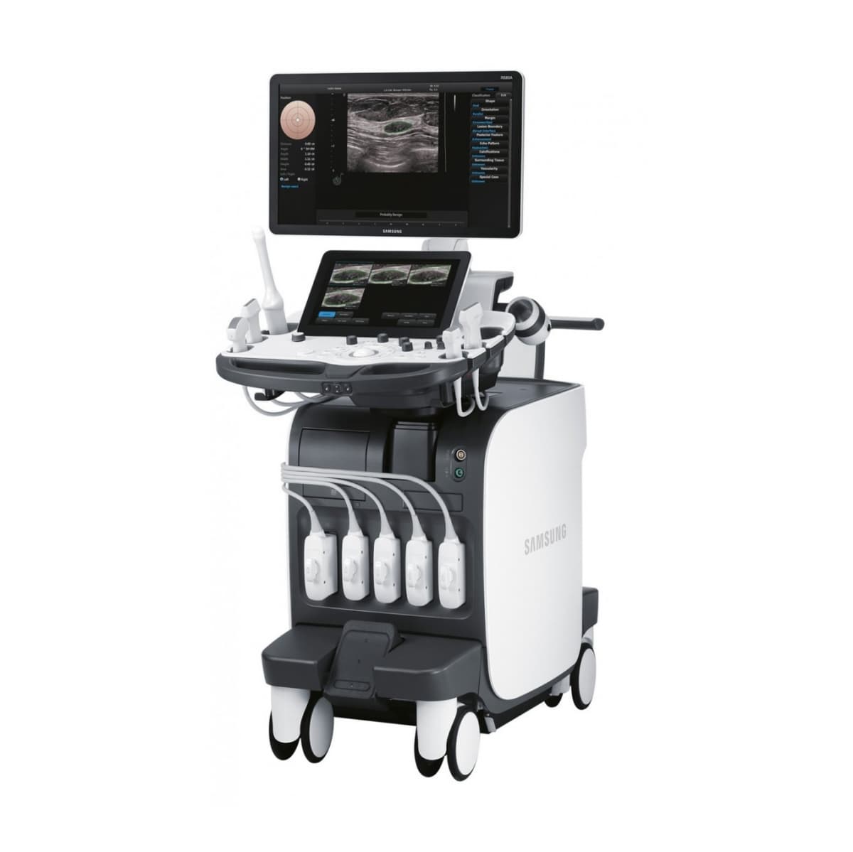
RS85 Prestige


RS85 Prestige
A Revolutionary Change inAdvanced Diagnostics
RS85 Prestige has been revolutionized with novel diagnostic features across each application based on the preeminent imaging performance. The advanced intellectual technologies are to help you confirm with confidence for challenging cases, while the easy-to-use system supports your effort involved in the routine scanning.
Especially, Samsung ultrasound's largest 27-inch OLED monitor enhances the diagnostic confidence of healthcare professionals by providing clear and stunning image quality.
Crystal ArchitectureTM
Crystal Architecture™, an imaging architecture that combines CrystalBeam™ and CrystalPure™,while based upon S-Vue Transducer™, is to provide crystal clear image.

PureVisionTM

PureVision™ is an image processing function that outputs with a good uniformity and clear image by performing Speckle Noise Suppression and Edge Enhancement on B-mode.
ShadowHDRTM

ShadowHDR™ selectively applies high-frequency and low-frequency of the ultrasound to identify shadow areas where attenuation occurs.
LumiFlow

LumiFlow™ ¹ is a function that visualizes blood flow in 3 dimensional-like to help understand the structure of blood flow and small vessels intuitively.
MV-FlowTM

MV-Flow™ ¹ visualizes microcirculatory and slow blood flow to display the intensity of blood flow in color.
EzHRI

HRI(Hepato Renal Index) is an index to quantify steatosis of a liver by comparing echogenicity between liver parenchyma and renal cortex. EzHRI™ ¹ places 2 ROIs on the liver parenchyma and renal cortex and provides HRI ratio.
S-Fusion™

S-Fusion™ enables simultaneous localization of a lesion using real-time ultrasound in conjunction with other volumetric imaging modalities. Samsung's Auto Registration helps quickly and precisely fuse the images, increasing efficiency and reducing procedure time. S-Fusion™ enables precise targeting during interventional and other advanced clinical procedures.
TAI™ ¹

TAI™ ¹(Tissue Attenuation Imaging) provides quantitative tissue attenuation measurement to assess steatotic liver changes.
HQ VisionTM

HQ-Vision™ provides clearer images by mitigating the characteristics of ultrasound images that are slightly blurred than the actual vision.
S-Detect™ for Thyroid

S-Detect™ for Thyroid ¹, ² analyzes selected lesions in the thyroid ultrasound study and shows the analysis data, provides standardized reporting based on the ATA, BTA, EU-TIRADS, K-TIRADS, and ACR TI-RADS guidelines; and helps diagnosis with the streamlined workflow.
S-Shearwave ImagingTM


Shearwave Imaging™ ¹ allows the noninvasive assessment of stiff tissues in various
applications. The color-coded elastogram, quantitative measurements, display options, and
user-selectable ROI functions are useful for accurate diagnosis.
S-Detect™ for Breast

S-Detect™ for Breast ¹, ² analyzes selected lesions in the breast ultrasound study and shows the analysis data, applies BI-RADS ATLAS* to provide standardized reporting; and helps diagnosis with the streamlined workflow.